-
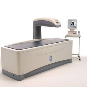
GE LUNAR Prodigy Bone Densitometer
View ProductSpecifications
Available Applications and Options
- AP Spine, Femur Dual, Femur Advance Hip Assessment with Hip Axis Length, Cross Sectional Moment of Inertia and Femur Strength Index Total Body*
- Body Composition* (with fat/lean assessment) Dual Energy Vertebral Assessment (DVA) Forearm Lateral Spine BMD
- Orthopedic Hip Analysis Pediatric*?Infant Total Body*** Small Animal
- One Vision, One Scan, Composer with 10-year Fracture Risk assessment Practice Management Report Dexter PDA interface software** Computer Assisted Densitometry (CAD) Tele Densitometry**?DICOM (Worklist – Color Print and Store)**?Multi User Data Base Access (3/10)**HL7 Bidirectional interface **
Software Platform
- Advanced intuitive graphical interface, Multiple Patient directories with Microsoft Access® database, Smart Fan TM for scan window optimization and dose reduction? Automated Scan mode selection ?Auto Analysis TM for a better precision
- Customized Analysis for clinical flexibility? Exam Comparison process? BMD or s BMD results (BMC and Area)?Extensive Reference Data
- > 12,000 subjects – NHANES and several Regional Lunar Reference Data User defined Reference Population
- T-score, Z-score, % Young-Adults and % Age-Match Automated WHO Background evaluation Patient trending with previous exam importation Multiple languages available
- Multimedia Online Help
Typical Scan Time and Radiation Dose at the best Precision
- AP Spine : 30 sec : 0.037mGy (< 1%CV)?Femur : 30 sec : 0.037 mGy (< 1%CV)?Total Body/ Body Comp. : 4 min 30sec: 0.0003 mGy (< 1%CV)
Calibration and Quality Assurance
- Automated test program with complete mechanicals and electronics tests and global measurement calibration Automated QA Trending with complete storage
Scanning Method
- Narrow Fan Beam (4,5° angle) with Smart Fan, MVIR and Tru View algorithms
X-ray characteristics
- Constant potential source at 76kV Dose efficient K-edge filter
Detector technology
- Direct-Digital CZT (Cadmium Zinc Telluride) detector Energy sensitive solid state Array
Magnification
- None – Object-plane measured
Dimensions (L x H x W) and weight
- 263 x 111 x 128 cm – 272 kg (Full)?202 x 111 x 128 cm – 254 kg (Compact) Vinyl table pad
External shielding
- Not required : X-ray safety requirements may vary upon destination.
Environnemental requirements
- Ambient temperature: 18-27°C?Power: 230/240 VAC ±10%, 10A, 50/60 Hz Humidity: 20% – 80%, non-condensing
Computer workstation
- Windows XP® Professional Intel processor computer, printer and monitor
-
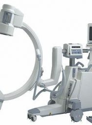
GE OEC 9000
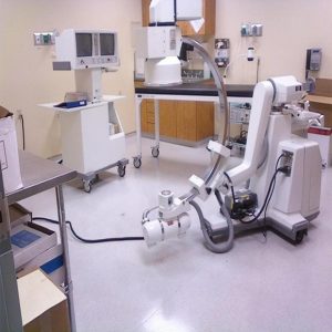
View ProductThe original c-arm that launched the OEC legend, the GE OEC 9000 remains a multiple application mobile x-ray imaging system that is flexible enough for use in general surgery, orthopedic surgery, trauma, and pain management.
Features
9000 C-Arm
Dual 17″ Monitors, 6″/9″ Image Intensifier
60 Image Storage, Printer, Food Switch
LIH Last Image Hold, Operator's Manual, 120V, 60Hz
2.5 kHz high frequency, 6.0 kW full-wave generator
Pulsed fluoro capability
Rotating anode x-ray tube
Digital Subtraction Angiography
Gamma correction
Dynamic real-time averaging
Motion Artifact Reduction
Expansion packages
Applications
General Surgery, Interventional Procedures, Neurology, Neurosurgery, Orthopedic, Pain Management, Trauma, Vascular.
-
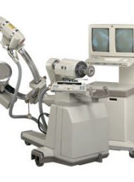
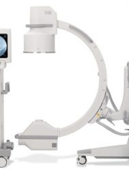
GE OEC 9400 C ARM
View ProductDual Monitors
Tri-Mode 4.5″/6″/9″ Image Intensifier
60 Image Storage
Rotating anode
Digital image enhancement (windowing)
Variable edge enhance(track ball)
Fluoro Boost
Sharpen Function
Gamma Correction
MARS-Motion Artifact Meduction
Sixty-image digital storage.
Video tape recorder (VTR)
LIH-Last Image Hold
-
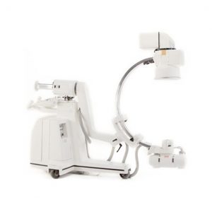
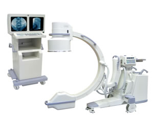
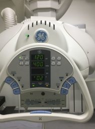
GE OEC 9800 Plus Features
View ProductSmartView pivot joint
SmartMetal
SmartWindow
AutoTrak
Pulse fluoro, High Level Fluoro and Digital Cine modes
Low dose features
15 kW generator
Rotating anode X-ray tube
Patented battery buffer technology
Upgradable to 16-bit image processing to deliver 1k x 1k images
Upgradable to 8 fps or 15 fps with real-time subtraction
Applications
General Surgery, Orthopedic, Pain Management, Peripheral Vascular, Spinal
-
GE Proteus Rad Rooms
View ProductThe most sought after used radiographic system.
The GE Proteus is a simple yet reliable radiographic system with overhead tube, elevating table, and bucky wallstand. This system includes phototiming, auto-collimation, and programmable APR’s. Techniques and image receptors are selectable at the tube head. Installation, warranty, and refurbishment are available.
Specifications:
Jedi high-frequency generator with programmable APR’s, touch screen LCD console, and self-calibrating reliability.
Elevating table with 4-way floating tabletop. Includes variable SID, auto-collimation, photo-timing, oscillating grid, and foot pedal controls.
Overhead tube crane with automatic SID detenting, auto-collimator, technique selection, image receptor selection, and variable vertical SID.
Bucky wall stand includes photo-timing and cassette sizing for auto-collimation.
-
GE Proteus X Ray 2012
View ProductThe Proteus XR/a is a full-featured radiography system designed to address nearly any clinical need. As a general purpose radiographic system, the Proteus XR/a offers broad clinical flexibility, excellent image quality, dose efficiency, ease of use, and outstanding reliability, all at an affordable price.
- Elevating, 4-Way Float Table
- Front and Rear Positioning Pedal Controls with Safety Lock
- Anti-Collision Sensor with Alarm
- LCD Color High Resolution Touch Screen
- Jedi Self-Calibrating Generator
- Three Phase
- Anatomical programming
- Auto Collimators: Inboard Bridge with Proteus OTS (Overhead Tube Support),
- Proteus Wall Stand with AEC (Auto Exposure Control)
- 80 kW High Frequency Generator
- 150 kVA
- 100 Amp Max
- 125 kV
- 600 mA
- Multi-size cassettes
-
GE RFX systems
View ProductFeatures
Table Features:
Quiet angulation drive with soft start and stop feature.
Tabletop longitudinal drive interlocked with angulation drive to prevent collision with floor and ceiling.
Tabletop and footrest extend downward toward floor with table vertical.
Range of Motion:
73.5” (187 cm) fluoroscopic beam center-to-floor dimensions when table is vertical for cervical esophagus coverage on 6′ 8” (2.0 m) tall patients.
11” (28 cm) transverse fluoroscopic carriage travel.
20” (50 cm) maximum caliper opening between bottom of spotfilmer or pedestal and tabletop. 19” (48 cm) maximum with 4-way flat top.
Structural plastic front and end panels shaped to eliminate sharp edges and barium traps while resisting strains, dents and corrosion.
Undertable X-ray tube equipped with a forced oil/ air heat exchanger for improved cooling.
Convenient Operator Features:
All table controls needed by technologist are located conveniently on table.
Interlocked patient step eliminates need for accessory foot stool.
Patient hand grips may be quickly attached at any point on front or rear of tabletop.
Myelographic Stop:
When automatic stop at horizontal is defeated for myelography and other needle intensive procedures, the compression cone drive is disabled.
Efficient Bucky Design:
Entrance type, three field Quantamat lonization Bucky Detector designed for minimum object film distance.
HGrid lines virtually invisible at 8 msec with 103-line grid.
RFX 88 Spotfilm Device:
Accepts 9.5” x 9.5” (24 x 24 cm) cassette sizes for operating simplicity and rapid access from fluoro mode.
Effort sensing, longitudinal power assist control in positioning handle.
1-on-1, 2-on-1 longitudinal, 2-on-1 transverse, and 4-on-1 formats.
Spotfilm access time from fluoro mode is approximately 0.75 seconds.
Rapid-sequence spotfilms are accomplished with approximately 0.5 seconds between exposures.
Table Bucky “center-finder” feature.
-
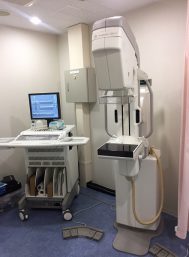
GE SENOGRAPH 2000D DIGITAL MAMOGRAPHY
View ProductREFURBISHED BY GE 2010
Full Digital Mammography. with RWS – Review Workstation and ICAD
LowNoise Detectors and High DQE; Single-Phase High Frequency; 22-49 kV Range; mAs Range 4-600; Automatic Exposure Detector; Parameters Control @ kVp, Track, Filter and mAs. Digital Detector type aSi – Csi; Imaging Area 19.2 cm x 23 cm; Acquisition Computer Sun Ultra 10; 21” Monitor; 20gB Hard Disk Capacity; DICOM (Print, STRG, Commit, Query – Retrieve, Modality Worklist) Molybdenum and Rhodium Rotating; Focal Spots 0.15 and 0.3; AutoCollimation; Flat Panel 19.2cm X 23cm Digital; Electromagnetic Locks; 66cm SID; Radiation Shield; Manual and Motorized Compression with Programmable Speed. Complete set of compression paddles, magnification platform and patient ID.
Composed of 1 Gantry, 1 Generator / Conditioner Cabinet, 1 Small Control Console, Glass Shield, 1 AWS Station with Monitor, Manuals, Magnification and Spot Paddle and 2 Regular Compression Paddles.
-
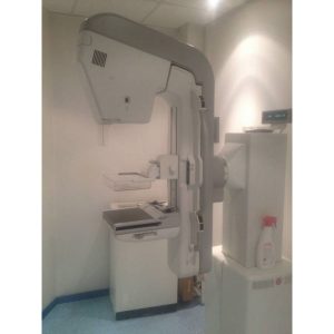
GE SENOGRAPH DMR PLUS 2002
View ProductIncludes all standard accessories plus the following:
- Compression paddles
- Includes Two Patient Bucky
- 18×24
- 24×30
- Magnification Paddles
- Biopsy
- 2 cassette Holders
- 2 Cassettes – 1 each size
- Schematics
- Technical Manual
- Operators Manual
- Service Kit


