-
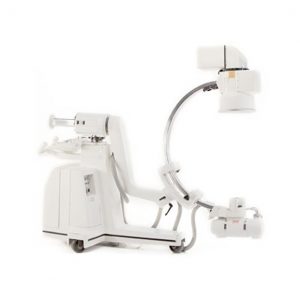
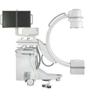
GE OEC 9400 C ARM
View ProductDual Monitors
Tri-Mode 4.5″/6″/9″ Image Intensifier
60 Image Storage
Rotating anode
Digital image enhancement (windowing)
Variable edge enhance(track ball)
Fluoro Boost
Sharpen Function
Gamma Correction
MARS-Motion Artifact Meduction
Sixty-image digital storage.
Video tape recorder (VTR)
LIH-Last Image Hold
-
GE OEC 9000
View ProductThe original c-arm that launched the OEC legend, the GE OEC 9000 remains a multiple application mobile x-ray imaging system that is flexible enough for use in general surgery, orthopedic surgery, trauma, and pain management.
Features
9000 C-Arm
Dual 17″ Monitors, 6″/9″ Image Intensifier
60 Image Storage, Printer, Food Switch
LIH Last Image Hold, Operator's Manual, 120V, 60Hz
2.5 kHz high frequency, 6.0 kW full-wave generator
Pulsed fluoro capability
Rotating anode x-ray tube
Digital Subtraction Angiography
Gamma correction
Dynamic real-time averaging
Motion Artifact Reduction
Expansion packages
Applications
General Surgery, Interventional Procedures, Neurology, Neurosurgery, Orthopedic, Pain Management, Trauma, Vascular.
-
GE Stenoscop 1991 C-Arm
View ProductFlouro Boost
Vascular
DSA
Roadmapping
Dual 17″ Monitors, 6″/9″ Image Intensifier,
100 Image Storage, Printer, Food Switch,
Hand Switch, LIH Last Image Hold,
120V, 60Hz
Operator's Manual
-
GE OEC 9800 Plus Features
View ProductSmartView pivot joint
SmartMetal
SmartWindow
AutoTrak
Pulse fluoro, High Level Fluoro and Digital Cine modes
Low dose features
15 kW generator
Rotating anode X-ray tube
Patented battery buffer technology
Upgradable to 16-bit image processing to deliver 1k x 1k images
Upgradable to 8 fps or 15 fps with real-time subtraction
Applications
General Surgery, Orthopedic, Pain Management, Peripheral Vascular, Spinal
-
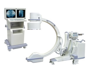
GE Fluorostar 7900 c-arm
View Product“Compact and modular,” these words describe the GE Fluorostar 7900 c-arm best. It is a high quality imaging device for use in the operating room, emergency room, intensive care unit, doctor’s offices, or anywhere else it could squeeze into. Its small size and easy maneuverability allows it to enter and fit in tight spaces. It comes with either single or dual 19-inch flat screen monitors, eliminating the need to wheel around a separate cart for viewing devices. It offers the combination of a high-resolution CCD camera and a 1k x 1k image processing capability.
Its modular design allows for versatility by extending the unit’s clinical capabilities. A Cine module enhances acquisition frame rates to as high as 25 frames per second; this is good for vascular procedures. The vascular module enables the unit to perform subtraction, peak opacification, and roadmapping. It pulsed modes and reduced dose processing decrease both the patients’ and staff’s exposure to radiation.
With advantages such as low radiation, superior imaging capabilities, and easy transport and positioning, the GE Flurostar 7900 c-arm may be the imaging unit your practice needs.
GE Fluorostar 7900 Features
- Touchscreen interface
- 1k x 1k resolution
- 540-image storage per acquisition
- Modular expansion options
- Up to 25 fps acquisition rates
- Integrated DICOM
- Hard drive for image storage of up to 60,000 images
- Single or dual 19-inch flat screen monitors
- USB support
- Image export to CD/DVD
Applications
Cardiac, Emergency Medicine, Endoscopic, Intensive Care, Neurology, Orthopedic, Urology, Vascular.
-
ACHILLES BONE ULTRASONOMETER
View ProductCERTAINTY
Proven in 11 prospective clinical studies.
High negative predictive value
Dynamic signal-strength compensation allows measurement over a wide range of bone densities
Bi-directional measurement helps ensure a consistent reading
Controlled measurement of site temperature for greater accuracy than other QUS systems
Results expressed as Stiffness Index—a composite of Speed of Sound (SOS) and Broadband Ultrasound Attenuation (BUA)—which compensates for the effect of heel width and temperature
Real-time image provides visual confirmation of proper heel placement (Achilles InSight only)
COMFORT
No messy gels
Warm, water-filled membranes hug the heel
Large, easy-to-read color display tilts towards the patient
No ionizing radiation
Quick exam—just a few minutes from shoe off to shoe on
CONVENIENCE
No special rooms
Durable and easily portable
Generates full-sized and full-color reports
On-board memory stores up to 2,000 patient records—no need to carry a computer
Customizable measurement workflows allow you to select which patient data to collect and report
-
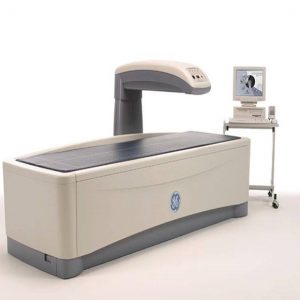
Hologic Discovery A
View ProductThe Discovery dual energy X-ray absorptiometry (DXA) system provides a single comprehensive platform for the assessment of three major health issues: osteoporosis including vertebral fracture assessment, cardiovascular disease, and obesity.
Hologic Discovery is Designed with Efficient Workflow in Mind!,The evaluation of bone health has become an essential component of your patients’ wellness. Hologic Discovery, our most advanced QDR Series bone densitometer, combines the proven clinical value of Bone Mineral Density (BMD) measurement and vertebral fracture assessment. Hologic pioneered the integration of BMD measurement with Instant Vertebral Assessment (IVA) allowing point-of-care assessment of the two most definitive factors associated with osteoporotic fracture risk: low bone mineral density and the presence of vertebral fracture. BMD and IVA are both powered by our exclusive One Pass linear fan-beam technology improving the accuracy, speed and convenience of assessing osteoporosis. ?Discovery’s technology innovations raise the standards One Pass Technology, Single sweep scanning enables superior image quality and unparalleled precision test after test, year after year ?Vertebral Fracture Assessment ?Recognized as the standard in fracture risk assessment is now even better with our High Definition Instant Vertebral Assessment, CAD fx, Computer aided fracture assessment provides physicians with tools to automatically recognize fractures ?Comprehensive Reporting Solutions. Hologic leads the industry in with reporting solutions for bone densitometry that streamline workflow and meet your reporting needs, Connectivity Solutions, Hologic Discovery connectivity solutions enable seemless communication to streamline workflow and increase productivity
Discovery delivers:
Exceptional Precision and Accuracy
High Definition Digital DXA Detectors – To improve fracture detection and to visualize abdominal aortic calcifications1.
Speed and Image Quality – Discovery imaging technology captures the hip and spine with as fast as 10-second regional scanning time.
Superb Visualization1 – The Discovery system’s exclusive design utilizes a high resolution detector array paired with true fan-beam linear acquisition geometry.
Consistency from Exam to Exam – The Discovery system performs continuous, automatic calibration, ensuring precise measurements results from exam to exam.
All Discovery DXA systems are powered by our exclusive OnePass technology. OnePass single sweep scanning delivers superb image quality1 and precision2. Our exclusive design utilizes a multi-element digital detector array paired with true fan-beam acquisition geometry, enabling rapid, dual-energy bone density measurements. OnePass scanning is designed to eliminate beam overlap errors and image distortion found in rectilinear acquisition techniques resulting in superb image quality and data stability.
- Radiological Vertebral Assessment
- Forearm, Dual Hip
- 10 Second Femur
- 10 Second Lumbar
- IVA
- Linear X-Ray Fan Beam Utilizing Motorized Table and C-Arm
- Computer Worktable with PC, Monitor
- Printer
- CD-RW Drive
- Network Ready
-
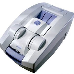
GE LUNAR Prodigy Bone Densitometer
View ProductSpecifications
Available Applications and Options
- AP Spine, Femur Dual, Femur Advance Hip Assessment with Hip Axis Length, Cross Sectional Moment of Inertia and Femur Strength Index Total Body*
- Body Composition* (with fat/lean assessment) Dual Energy Vertebral Assessment (DVA) Forearm Lateral Spine BMD
- Orthopedic Hip Analysis Pediatric*?Infant Total Body*** Small Animal
- One Vision, One Scan, Composer with 10-year Fracture Risk assessment Practice Management Report Dexter PDA interface software** Computer Assisted Densitometry (CAD) Tele Densitometry**?DICOM (Worklist – Color Print and Store)**?Multi User Data Base Access (3/10)**HL7 Bidirectional interface **
Software Platform
- Advanced intuitive graphical interface, Multiple Patient directories with Microsoft Access® database, Smart Fan TM for scan window optimization and dose reduction? Automated Scan mode selection ?Auto Analysis TM for a better precision
- Customized Analysis for clinical flexibility? Exam Comparison process? BMD or s BMD results (BMC and Area)?Extensive Reference Data
- > 12,000 subjects – NHANES and several Regional Lunar Reference Data User defined Reference Population
- T-score, Z-score, % Young-Adults and % Age-Match Automated WHO Background evaluation Patient trending with previous exam importation Multiple languages available
- Multimedia Online Help
Typical Scan Time and Radiation Dose at the best Precision
- AP Spine : 30 sec : 0.037mGy (< 1%CV)?Femur : 30 sec : 0.037 mGy (< 1%CV)?Total Body/ Body Comp. : 4 min 30sec: 0.0003 mGy (< 1%CV)
Calibration and Quality Assurance
- Automated test program with complete mechanicals and electronics tests and global measurement calibration Automated QA Trending with complete storage
Scanning Method
- Narrow Fan Beam (4,5° angle) with Smart Fan, MVIR and Tru View algorithms
X-ray characteristics
- Constant potential source at 76kV Dose efficient K-edge filter
Detector technology
- Direct-Digital CZT (Cadmium Zinc Telluride) detector Energy sensitive solid state Array
Magnification
- None – Object-plane measured
Dimensions (L x H x W) and weight
- 263 x 111 x 128 cm – 272 kg (Full)?202 x 111 x 128 cm – 254 kg (Compact) Vinyl table pad
External shielding
- Not required : X-ray safety requirements may vary upon destination.
Environnemental requirements
- Ambient temperature: 18-27°C?Power: 230/240 VAC ±10%, 10A, 50/60 Hz Humidity: 20% – 80%, non-condensing
Computer workstation
- Windows XP® Professional Intel processor computer, printer and monitor
-
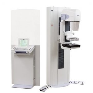
Lorad M-IV Platinum Mammography
View ProductBi-angular X-ray Tube
- Custom design with high speed anode rotation
- significantly higher mA loading and output
- Maintain tight focal spot size tolerances
- Eliminate the need to wait for the tube to cool down
- Receive exceptionally high quality, high resolution images for both full field and magnification viewsFAST Paddle
- Fully Automatic Self-Adjusting Tilt (FAST) Paddle
- Improve imaging
- Provide greater patient comfort
- Ensure more uniform compression across the entire breast
- Superior image quality
- Improve immobilization of breast tissue
- Reduce motion artifacts
- Prevent loss of compression or over-compression at the chest wall
- Enhance patient comfort and optimal imagingAdvanced Solutions For Breast Imaging
- Offer a full compliment of diagnostic accessories
- Obtain flexibility and functionality essential for the diagnostic evaluation of an area of interest
- Quickly convert the M-IV to an advanced imaging system for stereotactic, needle localization and ultrasound procedures with specialized accessories
- Perform exams while under compression
- Visualize the breast tissue in the same plane as the mammogram
- Increase diagnostic assurance without any inconvenience to your patients
Ease Of Operation
- Advanced automated features
- Get outstanding consistency and operator efficiency
- AutoFilter
- Conveniently select appropriate exposure and filtration settings
- Automatic Collimation Complete
- Eliminate manual apertures
- Four Exposure Modes
- From fully automatic to totally operator-selected, based on operator preference
- Operator Preferences
- Store and automatically activate upon log-inImage Quality
- Capture high quality images
- High Transmission Cellular Grid (HTC Grid)
- Get higher contrast images
- Bi-angular X-ray Tube
- Enhance magnification views with a custom designed high performance tube
- FAST Paddle
- Exclusive Fully Automatic Self-adjusting Tilt Paddle
- Achieve more uniform compression
- 3-Cell, 7 Position AEC Sensor
- Unique Automatic Exposure
- Control for more precise technique selection
Gantry:
Height: 75 in
Width: 25 in
Depth: 50 inWeight: 850 lbs
Console With Lead Glass Shield:
Height: 74 in
Width: 32 in
Depth: 17 inWeight: 250 lbs
Generator
High frequency constant potential
Ma range (IMAGING SYSTEM)
30, 100Kv range (X-RAY GENERATOR) 20increments of 1 kV
mAs RANGE (X-RAY TUBE) 3
Power Needed
200-240 VAC 10%, 35 A, 50/60 Hz2
-
GE SENOGRAPH DMR PLUS 2002
View ProductIncludes all standard accessories plus the following:
- Compression paddles
- Includes Two Patient Bucky
- 18×24
- 24×30
- Magnification Paddles
- Biopsy
- 2 cassette Holders
- 2 Cassettes – 1 each size
- Schematics
- Technical Manual
- Operators Manual
- Service Kit
-
GE SENOGRAPH DS DIGITAL MAMMOGRAPHY UNIT
View ProductAWS V3 Software Version 53.10.1
Inventory
Gantry with Interconnect Cables
Generator with Chiller
Acquisition Workstation with Radiation Shield
UPS
Monitor
ADS Computer
IDC Computer
Keyboard and Mouse
Control Console
Bucky
2 Footswitches
8 Compression Paddles
2 Magnification Platforms
Face Guard
Accessory Cabinet
Dosimeter Support Bracket
Flat Field Phantom
IQST Phantom
AOP Plates
Chiller Kit
Detector Defect Map Disk
Software
Manuals
-
2006 SIEMENS FULL FIELD DIGITAL MAMMOGRAPHY
View ProductNOVATION DR with RWS/Review Doctors station.
Year 2006 Full Field Digital Mammography, Microprocessor High Frequency generator; 23-35 kV Range; mAs Range 25-188; Automatic Exposure Detector; Parameters Control @ kVp, Track, Filter and mAs. AEC Detector; Digital Detector: aSe. Imaging Area 24 cm x 29 cm (Large Detector), DICOM. Acquistion Workstation with SYNGO Operating System. Revision VA20E., Focal Spots 0.1 and 0.3; AutoCollimation; Flat Panel 19” TFT Color Display; 65cm SID; Manual and Motorized Compression with Programmable Speed. Complete set of compression paddles.


