-
-
-
-
-
-
-
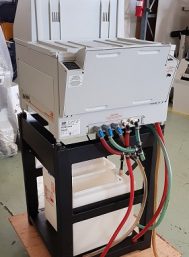
AFP Mini Medical 90 Developer
View ProductAFP Mini Medical 90 Developer
The Mini-Medical/90 is a perfect choice for small offices, clinics, satellite health care facilities and mobile installations. Compact, reliable and easy to use, the Mini-Medical delivers high quality x-ray images, week after week, year after year. A full warranty will help your practice operate worry-free for years.
Characteristics:
- Built-in Quality Control Indicators
- Automatic Chemical Replenisher
- No Plumbing Necessary (optional kit)
- Automatic Easy Water Control
- Infrared Dryer
- Space Saving Size
- Reliable Efficiency
- Easy to Install, Use, & Maintain
-
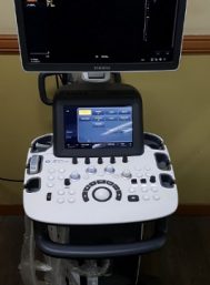
Ultrasonido Samsung UGEO H60 2015
View ProductLa tecnología aplicada en el sistema UGEO H60 incluye:
TDC Digital: El UGEO H60 es el primer ultrasonido de gabinete que cuenta con esta innovación, lo que brinda un manejo mas rápido e intuitivo del mismo.
autoIMT: Herramineta de software especializada capaz de medir automáticamente el espesor de las capas de la intima –media.
SDMR: Herramienta de software que brinda imágenes en calidad de resonancia magnética.
BEAMSteering: Herramienta de software capaz de resaltar la aguja en procedimientos como el bloqueo de nervios, con el fin de poder realizar un estudio mucho mas preciso en menor tiempo.
Características:
18.5″ LED/HD monitor with Articulating Arm, 10.1″ wide touch screen, 4 probe ports, Independent steer & lockable wheels, User customizable touch menus, User customizable keys, QuickScan, Digital TGC presets (Cardiac, OB/GYN, Vascular/Urology), Live 3D/4D, Hybrid Imaging Engine, SDMR, S-Flow, SCI, Auto Image Optmization, Auto NT Measurement, Auto IMT, Automated B/M/D Measurement ,Harmonic imaging
Trapezoidal Imaging, Panoramic Imaging, Write Zoom, Speckle Reduction, TDI [Tissue Doppler Imaging]
THI [Tissue Harmonic Imaging], Strain-based Elastography, Volume NT&IT, 3D XI, Beam Steering, SmartExam / Ease Protocol, DICOM 3.0, DICOM SR OB/GYN, JPEG, WMV, & AVI output, USB Port ,Solid State Drive storage technology, DVD/CD RW, Flexible ReportTransductores:
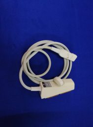
- Transductor Convexo 3D VE4-8 [ 4 – 8 MHz ]
- Transdcutor Convexo C2-8 [ 2 – 8 MHz ]
- Transductor Endocavitario EVN4-9 [ 4 – 9 MHz]
- Impresora papel Sony B/N
-
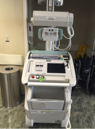
CR X RAY FUJI FCR GO
View Product2009 Fuji FCR GO Portable Digital X-Ray System Model: FCR-MB 101 DOM: 2009
- Computed Radiography Model
- CR-IR 358RU
- X-Ray Collimator Type: ZU-L3SA
- X-Ray Tank Unit Type M-5CE-30
- X-Ray Tube Type: R-5CE-30
- Digital * Built-In CR Reader
- Touch Pad Electronic Controls
- Dynamic Visualization
- Manual Collimator
- Rotating Vertical Column
- Telescopic Tube Arm
- Hand Switch
- Cassette Drawer
- Cassettes: (1) 10×12, (2) 14×17 Electromechanical Brakes
-
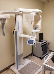
Planmeca ProMax® 2D X-ray
View ProductPlanmeca_ProMax_brochure_EN
Planmeca ProMax® 2D X-rayunit provides a wide range of extraoral imaging modalities:
- panoramic imaging for dental arch
- maxillary sinus imaging
- temporomandibular joint imaging
- 2D linear tomography
- cephalometry
Revolutionary technology
The Planmeca ProMax® platform uses robotic SCARA technology to provide
utterly precise arm movements needed in digital rotational radiography
for maxillofacial imaging. The three-axis SCARA robot arm moves freely without
any mechanical restrictions, offering superior imaging capabilities for both
existing and future technologies.
Easy upgrades
An existing digital Planmeca ProMax can be easily upgraded to 3D unit and new
imaging programs can be added with software upgrades ensuring superior
maxillofacial radiographs every time and complying with future imaging needs.
-
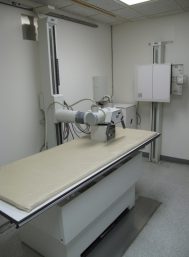
SUMMIT X RAY ROOM 2005
View Product- 300 mA, X Ray room Manucatured by Summit 2005
- CPI Generator 300 Ma 2005
- Wall to Table Columm
- 4 Way X Ray Table
- Wall Bucky
- 300 mA X Ray Tube
- Colimmator
- Summit Generator
- X Ray Transformer
- High Voltage Cable
- Service Manuals
-
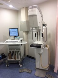
GE SENOGRAPH 2000D DIGITAL MAMOGRAPHY
View ProductREFURBISHED BY GE 2010
Full Digital Mammography. with RWS – Review Workstation and ICAD
LowNoise Detectors and High DQE; Single-Phase High Frequency; 22-49 kV Range; mAs Range 4-600; Automatic Exposure Detector; Parameters Control @ kVp, Track, Filter and mAs. Digital Detector type aSi – Csi; Imaging Area 19.2 cm x 23 cm; Acquisition Computer Sun Ultra 10; 21” Monitor; 20gB Hard Disk Capacity; DICOM (Print, STRG, Commit, Query – Retrieve, Modality Worklist) Molybdenum and Rhodium Rotating; Focal Spots 0.15 and 0.3; AutoCollimation; Flat Panel 19.2cm X 23cm Digital; Electromagnetic Locks; 66cm SID; Radiation Shield; Manual and Motorized Compression with Programmable Speed. Complete set of compression paddles, magnification platform and patient ID.
Composed of 1 Gantry, 1 Generator / Conditioner Cabinet, 1 Small Control Console, Glass Shield, 1 AWS Station with Monitor, Manuals, Magnification and Spot Paddle and 2 Regular Compression Paddles.


